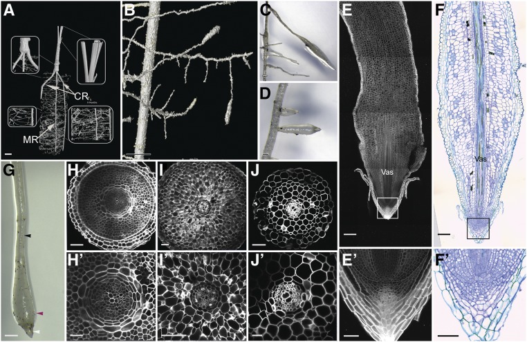
Figure 9.
Characterization of Date Palm Pneumatophores.
(A) and (B) XµCT images showing the root system architecture of the date palm: the primary root with secondary lateral horizontal growth and aerial roots with upward vertical growth. (B) is a zoomed image of (A).
(C), (D), and (G) Micrographs showing pneumatophores (n = 10).
(E) and (E′) Longitudinal sections of date palm pneumatophores stained with mPS-PI (n = 5).
(F) and (F′) DIC images of sections stained with toluidine blue O (n = 5).
(H) to (J′) Cross sections of different zones within the pneumatophore (n = 5); basal thin region (white arrowhead; see [H] and [H′]), basal thick region (purple arrowhead; see [I] and [I′]), and apical thin region (black arrowhead; see [J] and [J′]). (E′), (F′), (H′), (I′), and (J′) are enlargements of (E), (F), (H), (I), and (J), respectively. Images are representative of the total number (n) of roots that were studied. Bar in (A) = 20 mm; bar in (B) = 4.5 mm; bar in (C), (D), and (G) = 1 cm; bar in (E) and (F) = 100 µm; bar in (H), (I), (J), (E′), and (F′) = 50 µm; and bar in (H′), (I′), and (J′) = 25 µm. CR, crown root; MR, main root.

