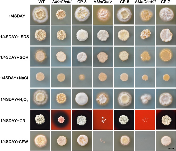
Fig 2. Colony morphology of the wild-type strain and the ΔMaChsIII, ΔMaChsV, ΔMaChsVII mutants.
Colony morphology of the MaChs deletion mutants on 1/4 SDAY or 1/4 SDAY supplemented with 0.1% SDS (sodium dodecyl sulfate), 1.5 mol l-1 Sorbitol, 0.5 mol l-1 NaCl, 500 μg ml-1 CR (Congo red), 50 μg ml-1 CFW (calcofluor white), 6 mmol l-1 H2O2 at 28°C. The fungal colonies were photographed after 5 d of incubation. Bar scale = 0.5 cm.

