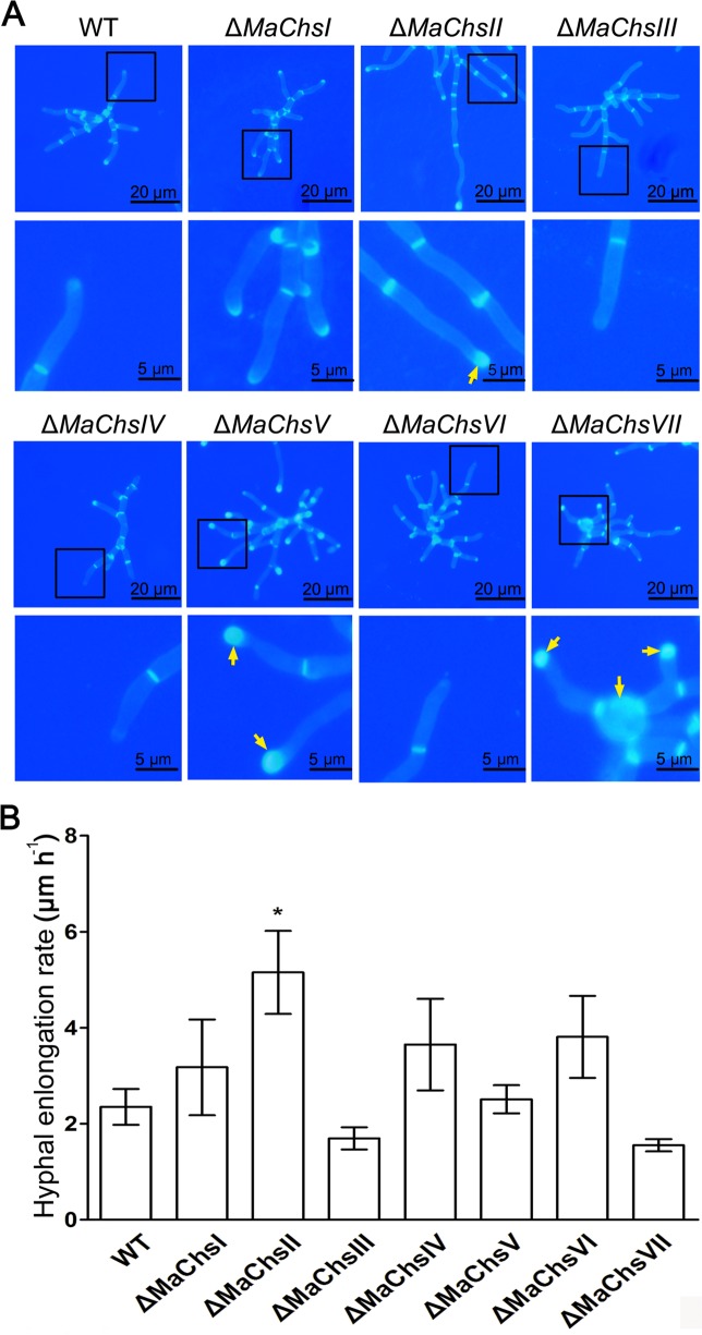
Fig 3. Growth and hyphal morphology of Chs mutants.
(A) Hyphal morphology of wild type and ΔMaChs mutants on 1/4 SDAY. Cell wall chitin of wild type and ΔMaChs mutant hyphae were observed staining with calcofluor white on a cover glass by fluorescence microscopy. Arrows indicate the site of chitin accumulation. (B) Hyphal growth rates of wild type and ΔMaChs mutants on 1/4 SDAY. A single asterisk above bars denotes significant difference, P < 0.05. Error bars indicate standard errors of three trials.

