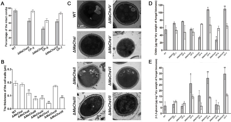
Fig 5. Cell wall structure and composition of wild type and ΔMaChs mutants.
(A) Measurements of cell wall integrity for the wild type and ΔMaChsIII, ΔMaChsV, ΔMaChsVII mutants. (B) The cell wall thickness of the wild type and MaChs mutants. (C) Transmission electron micrographs (TEM) images for the cell wall distortions seen in conidia produced by the wild type and MaChs mutants. Bar scale = 1 μm. (D) Chitin content as determined by acid hydrolysis of fungal cell wall. (E) β-1,3-glucan determined via quantification of the alkali-insoluble fraction of the cell wall. Samples were vegetative hyphae harvested from 3-day-old liquid 1/4 SDY cultures. A single asterisk above bars denotes significant difference, P < 0.05; double asterisks above bars denote significant difference, P < 0.01. Error bars indicate standard errors of three trials.

