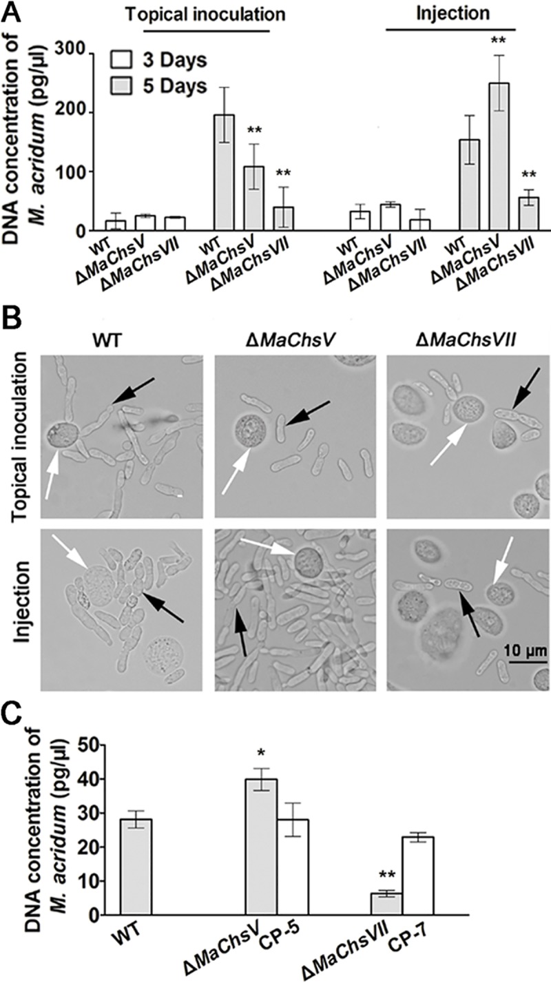
Fig 8. Fungal growth in the insect hemolymph and in vitro.

(A) Quantification of fungal growth. Total fungal DNA corresponding to the wild type, ΔMaChsV and ΔMaChsVII mutants in the locust hemolymph after topical inoculation and intrahemocoel injection was determined over the indicated time course as detailed in the Methods section. (B) Representative images of M. acridum hyphal bodies in the locust hemolymph at 5 d post-treatment. Black arrows indicate hyphal bodies. White arrows indicate locust hemocytes. (C) Quantification of fungal growth in isolated locust hemolymph (in vitro). Total DNA concentrations of the wild type, ΔMaChsV and ΔMaChsVII mutants grown in isolated locust hemolymph in vitro. A single asterisk above bars denotes significant difference, P < 0.05; double asterisks above bars denote significant difference, P < 0.01. Error bars indicate standard errors of three trials.
