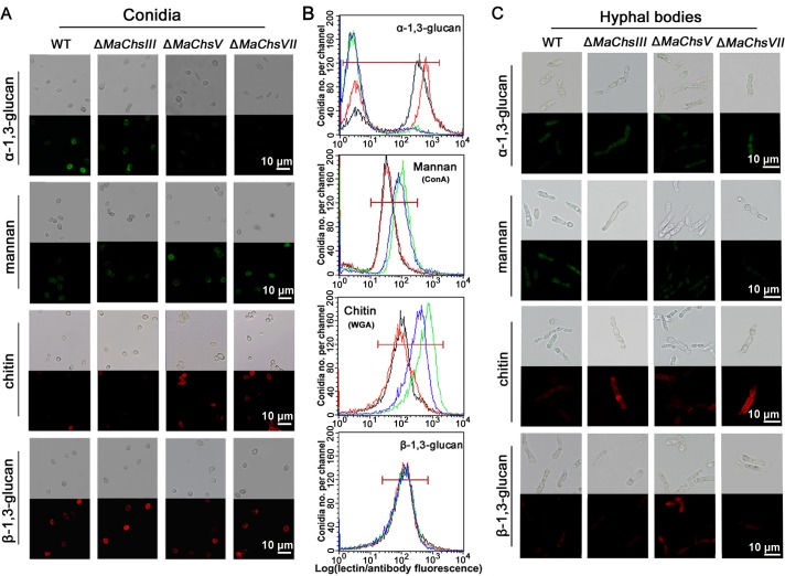
Fig 11. Detection of fungal cell wall surface carbohydrates.
(A) Detection of α-1,3-glucan, mannan, chitin, and β-1,3-glucan on conidial surface using fluorescent staining. (B) Representative flow cytometry results for distributions of lectin and antibody binding by M. acridum conidia from wild type (black line), ΔMaChsIII (red line), ΔMaChsV (green line), and ΔMaChsVII (blue line). (C) Detection of α-1,3-glucan, mannan, chitin, and β-1,3-glucan on hyphal body surface using fluorescent staining. The α-1,3-glucan on fungal cell wall surface staining by α-1,3-glucan-specific antibody, IgM clone MOPC-104E, with Alexa Fluor 488 goat anti-mouse IgG secondary antibody. Mannan on fungal cell wall surface staining by fluorescein-labeled ConA in fungal cell walls. Chitin on fungal cell wall surface staining by fluorescein-labeled WGA. The β-1,3-glucan on fungal cell wall surface staining by β-1,3-glucan-specific antibody with Alexa Fluor 594 goat anti-mouse IgG (H+L) secondary antibody.

