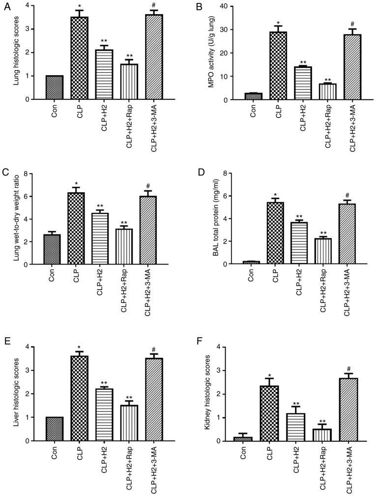
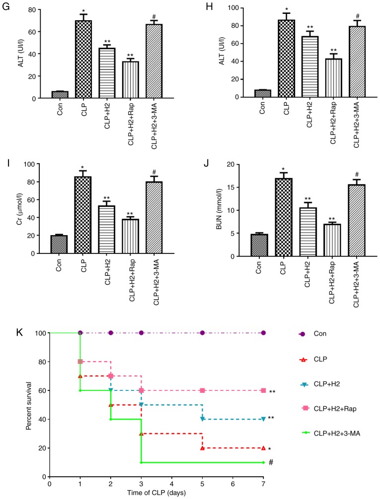
Figure 6.
Effect of autophagy-mediated NACHT, LRR and PYD domains-containing protein 3 inactivation on lung injury, biochemical parameters of liver and kidney and survival rate in LPS-induced macrophages treated with H2. Sepsis was produced by CLP. Septic mice were treated with H2, autophagy inducer Rap and autophagy inhibitor 3-MA. After 24 h, lung tissues were collected to detect (A) pathological tissue changes by hematoxylin and eosin staining, (B) MPO activity and (C) W/D weight ratio; (D) bronchoalveolar lavage fluid was collected to analyze total proteins. (E) Liver and (F) kidney tissues were collected to investigate pathological scores. Blood was obtained to measure the biochemical parameters (G) ALT, (H) AST, (I) Cr and (J) BUN. (K) Survival rate was analyzed at 1, 2, 3, 5 and 7 days post-CLP (n=20). Data are expressed as the mean ± standard deviation (n=6). *P<0.05 vs. Con group, **P<0.05 vs. CLP group, #P<0.05 vs. CLP+H2 group. LPS, lipopolysaccharide; CLP, cecal ligation and puncture; Rap, rapamycin; 3-MA, 3-methyladenine; MPO, myeloperoxidase, W/D, wet/dry; ALT, alanine transaminase; AST, aspartate transaminase; BUN, blood urea nitrogen; Cr, creatinine; H2, hydrogen; Con, control.


