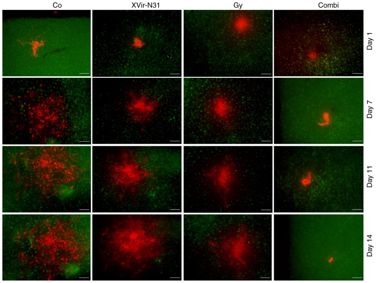
Figure 4.
Irradiation prior to XVir-N-31 infection delayed the growth of R28 tumor spheroids in human cortical brain slice cultures. R28mCherry spheroids were implanted in human brain slices stained with NeuO. The cultures were left either untreated (Co), irradiated with 3 Gy, infected with 5×10⁷ IFU XVir-N-31, or were irradiated with 3 Gy and infected using 5×10⁷ IFU XVir-N-31 24 h later. Images were captured every second day starting at the day of XVir-N-31 infection. Green staining represents NeuO staining in the brain slices. Red staining represents R28mCherry-expressing cells. Representative 1/6 spheres from 2 independent experiments is presented (scale bar=200 µm). IFU, infectious units; Co, control; Combi, 3 Gy tumor irradiation + an intratumoral XVir-N-31 injection (1.5×10⁸ IFU) 24 h later.

