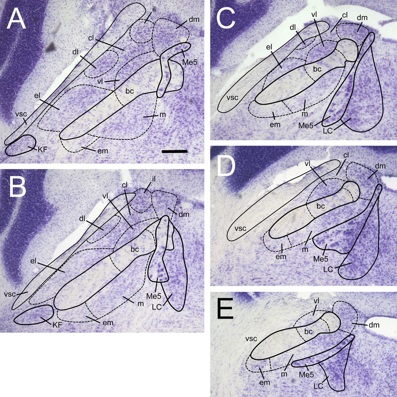
Figure 1.
Delineation of the PBN at 5 levels along the rostral-caudal axis of the mouse brain. Sections were nissl-stained with cresyl violet to reveal cytoarchitecture. This span of sections (40 μm thickness) is spaced out evenly over a 360 μm distance, starting at the most rostral section (A), approximately –5.24 mm relative to bregma. Sections are ordered A-E, from rostral to most caudal. PBN subnuclei are labeled as follows: dm, dorsal medial; m, medial; em, external medial; bc, brachium conjunctivum; vl, ventral lateral; cl, central lateral; dl, dorsal lateral; el, external lateral; il, internal lateral. All pictures at same magnification, scale bar in A = 100 μm. Other abbreviations are: vsc, ventral spinocerebellar tract; Me5, mesencephalic trigeminal nucleus; LC, locus coeruleus; KF, kolliker-fuse nucleus.

