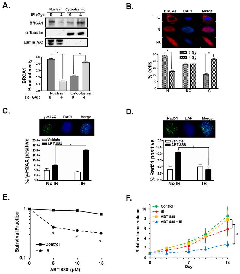
Figure 5. IR sensitizes glioblastoma cancer cells to PARPi through cytoplasmic translocation of BRCA1.
A, BRCA1 cytoplasmic translocation is measured by western blot of BRCA1 in the nuclear and cytoplasmic protein fractions of U87 cells treated with 0 or 4 Gy IR. α-tubulin was used as a cytoplasmic protein loading control while lamin A/C served as a loading control for the nuclear fractions. Bar graph summarizes the intensity of the BRCA1 band in the respective fractions from three independent experiments. B, representative images illustrate cytoplasmic (C), nuclear (N), or nuclear and cytoplasmic (NC) fluorescent immunohistochemical staining of BRCA1 (red). Nuclei are stained blue with DAPI. Bar graph illustrates the percentage of cells demonstrating nuclear (N), nuclear and cytoplasmic (NC), or cytoplasmic (C) staining of BRCA1 after treatment with 0 (grey bars) or 4Gy (black/white checkered bars). The result is the average of three experiments. C, representative photos illustrating fluorescent immunohistochemical staining of nuclear-γ-H2AX foci (green). Cell nuclei are stained blue with DAPI. Cells were pre-treated with 0 or 4 Gy IR, then treated 24h later with 0 or 10 µM ABT-888. After an additional 24h, cells were fixed, stained and assayed for γ-H2AX foci. Results are the summary of three experiments. D, representative photos illustrate fluorescent immunohistochemical staining of Rad51 foci (green). U87 cells were pre-treated with 0 or 4 Gy IR, then treated 24h later with 0 or 10 µM ABT-888 after an additional 24h cells were fixed and stained and assayed for Rad51 foci. Bar graph summarizes the percentage of U87 control (white bars) or ABT-888 treated cells (10 µM, black bars) with Rad51 foci after treatment with no radiation or 4h after treatment with 4Gy IR from three independent experiments. E, U87 cells were first treated with 0 (square, solid line) or 4Gy IR (circles, dashed line), 24h later the cells were treated with the indicated concentration of ABT-888 and the surviving cells were assayed by their ability to form colonies. Results summarize three independent experiments. F, U87 tumors were established in nu/nu mice and then subjected to no treatment (control, n = 6), treatment with a single dose of 4Gy IR (IR, n = 4), treatment with 25mg/kg ABT-888 daily for 5 days (ABT-888, n = 5), or a combination of 4 Gy IR followed by 5 daily treatments of 25mg/kg ABT-888 beginning 24h post-radiation (ABT-888 + IR, n = 6). *, p < 0.05.

