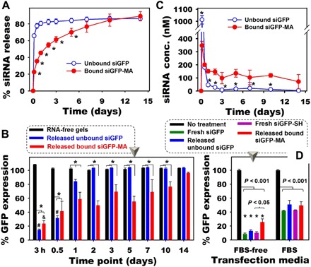
Fig. 3. Release of phototethered siRNA from the hydrogels and RNA bioactivity.

(A) Release profiles of siRNA (10 μg per 50 μl of gel) from 10% (w/w) DEX hydrogels into phenol red–free DMEM-HG (*P < 0.001 compared with unbound siGFP at the same time point). (B) GFP expression of HeLa cells treated with the same volume of releasates from different groups without the addition of transfection reagent and in the absence of FBS for 2 days (*P < 0.05; #P < 0.05 and &P < 0.05 compared to different time points of the same hydrogels). (C) Concentration of siRNA in releasate at different time points, which were used as transfection media to obtain (B) (*P < 0.05 compared with “bound siGFP-MA” at the same time point). (D) Bioactivity of released siGFP at the same siRNA concentration (350 nM) performed in DMEM-HG containing 0% FBS (“FBS-free”) or 2.5% (v/v) FBS [*P < 0.001 compared to the corresponding (color) FBS groups].
