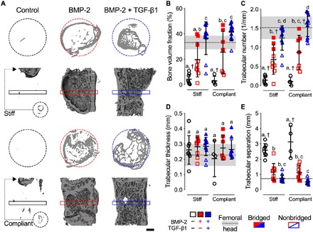
Fig. 2. Effects of morphogen priming of engineered mesenchymal condensations and in vivo mechanical loading on new bone quantity and architecture.

(A) Representative 3D microCT reconstructions, with bone formation illustrated at mid-shaft transverse (top) and sagittal (bottom) sections at week 12, selected on the basis of mean bone volume. Dashed circles show 5-mm defect region. Rectangular boxes illustrate transverse cutting planes. Note that, due to minimal bone regeneration, additional transverse sections for stiff and compliant no growth factor controls were derived from the proximal end of the defect (small dashed circles and arrows). Scale bar, 1 mm. (B) Morphometric analysis of bone volume fraction, (C) trabecular number, (D) trabecular thickness, and (E) trabecular separation at week 12 (n = 4 to 11 per group), shown with corresponding measured parameters of femoral head trabecular bone (n = 3; dotted lines with gray shading: means ± SD; †P < 0.05 versus femoral head). Individual data points are shown as means ± SD. Comparisons between groups were evaluated by two-way ANOVA with Tukey’s post hoc tests. Repeated significance indicator letters (a, b, and c) signify P > 0.05, while groups with distinct indicators signify P < 0.05.
