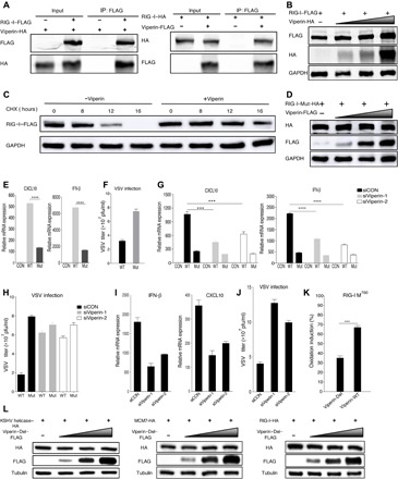
Fig. 6. Methionine oxidation catalyzed by viperin increases the stability and function of RNA helicase RIG-I.

(A) Reciprocal co-IP assays to examine physical interactions between viperin and RIG-I. (B) 293T cells were transfected with plasmids containing indicated genes. At 48 hours after transfection, WCLs were analyzed by immunoblotting. (C) 293T cells were transfected with plasmids containing indicated genes. At 24 hours after transfection, cells were treated with CHX (10 μg/ml). WCLs were then analyzed by immunoblotting. (D) 293T cells were transfected with plasmids containing indicated genes. At 48 hours after transfection, WCLs were analyzed by immunoblotting. (E) 293T cells were transfected with plasmids containing indicated genes. At 24 hours after transfection, cells were transfected with poly(I:C) for 16 hours. RNA was extracted, and cDNA was prepared to determine CXCL10 and IFN-β mRNA expression by qPCR analysis. (F) Reconstituted MEF cells were infected with VSV [multiplicity of infection (MOI), 0.1], and viral titer in the supernatant was determined by plaque assay. pfu, plaque-forming units. (G) 293T cells were transfected with plasmids containing indicated genes after transfection for 6 hours with siRNA, as indicated. At 24 hours after transfection, cells were transfected with poly(I:C) for 16 hours. RNA was extracted, and cDNA was prepared to determine CXCL10 and IFN-β mRNA expression by qPCR analysis. (H) Reconstituted MEF cells were infected with VSV (MOI, 0.1) after transfection for 24 hours with siRNA, as indicated, and viral titer in the supernatant was determined by plaque assay. (I) 293T cells were transfected with siRNA, as indicated. At 24 hours after transfection, cells were transfected with poly(I:C) for 16 hours. RNA was extracted, and cDNA was prepared to determine CXCL10 and IFN-β mRNA expression by qPCR analysis. (J) MEF cells were infected with VSV (MOI, 0.1) after transfection for 24 hours with siRNA, as indicated, and viral titer in the supernatant was determined by plaque assay. (K) Viperin wild type (Viperin-WT), viperin-Del, and RIG-I were purified separately from 293T cells. Oxidative reaction was performed in vitro, and methionine oxidized peptides were quantitatively determined by mass spectrometry analysis. (L) 293T cells were transfected with plasmids containing indicated genes. At 48 hours after transfection, WCLs were analyzed by immunoblotting. For (E to K), the data are expressed as means ± SEM; n = 3; ***P < 0.001 and ****P < 0.0001.
