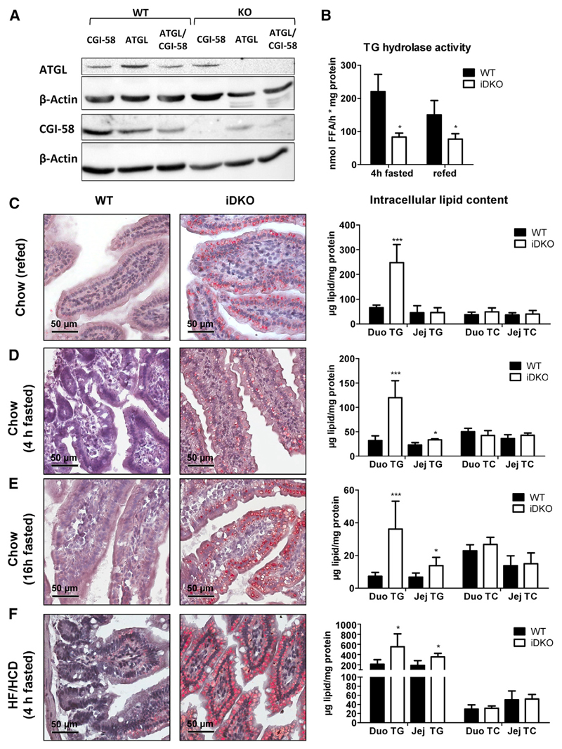
Figure 1. Loss of Intestinal ATGL and CGI-58 Leads to Augmented Intracellular Lipid Accumulation.
(A) Protein lysates (80 μg) were separated using SDS-PAGE, and protein expression levels of ATGL and CGI-58 in the jejunum of 4 h fasted, chow dietfed Atgl iKO, Cgi-58 iKO, and iDKO mice were determined using western blotting. Monoclonal anti-mouse β-actin served as loading control.
(B) TG hydrolase activity in the jejunum of chow diet-fed iDKO mice in 4 h fasted (n = 3 or 4) and refed states (12 h fasting, 2 h refeeding; n = 4 or 5).
(C–F) ORO staining of duodenal cryosections and biochemical quantification of intracellular lipid concentrations in chow diet-fed iDKO mice after refeeding (n = 4 or 5; C), fasted for 4 h (n = 3 or 4; D) or 16 h (n = 6–9; E), and in mice challenged with HF/HCD for 5 weeks and fasted for 4 h (n = 4 or 5; F). Data represent mean + SD. *p < 0.05 and ***p ≤ 0.001. Magnification, 40×; scale bar, 50 μm. Duo, duodenum; Jej, jejunum; TC, total cholesterol; TG, triglycerides.
See also Figure S1.

