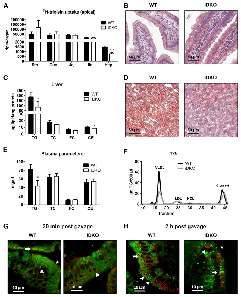
Figure 2. Intestinal Loss of ATGL and CGI-58 Ameliorates Hepatic Steatosis 30 min Post-gavage.
(A) Radioactivity in the SI and liver of chow diet-fed WT and iDKO mice (n = 4 or 5) 30 min post-gavage of 2 μCi [9,10-3H(N)]-triolein in 200 μL corn oil.
(B) ORO staining of duodenal sections 30 min after an oral lipid load.
(C and D) Biochemical (C) and histological (D) analysis of hepatic lipid levels 30 min post-gavage of 200 μL corn oil.
(E and F) Lipid concentrations (E) and (F) lipoprotein profiles in the plasma 30 min post-gavage of corn oil (200 μL). Data represent mean + SD (n = 3 or 4). *p < 0.05, **p ≤ 0.01, and ***p ≤ 0.001. Magnification, 40×; scale bar, 50 μm.
(G and H) Chow diet-fed mice were fasted for 16 h prior to an oral administration of 100 μL corn oil containing 1 μg/g body weight BODIPY FL C16 (green). Intestinal sections were co-stained with PLIN3 (red) to visualize colocalization with cLDs. PLIN3 immunofluorescence staining 30 min (G) and 2 h (H) post-gavage. Green background fluorescence results from high chlorophyll content in the chow diet (alfalfa). Arrows indicate cLDs originating from BODIPY-labeled FA, which colocalize with PLIN3; arrowheads indicate endogenous cLDs coated with PLIN3; stars indicate BODIPY-containing cLDs, which do not colocalize with PLIN3. Magnification, 100×; scale bar, 10 μm.
CE, cholesteryl esters; Duo, duodenum; FC, free cholesterol; HDL, high-density lipoprotein; Hep, hepar (liver); Ile, ileum; Jej, jejunum; LDL, low-density lipoprotein; PLIN3, Perilipin 3; Sto, stomach; TC, total cholesterol; TG, triglycerides; VLDL, very low density lipoprotein.
See also Figure S2.

