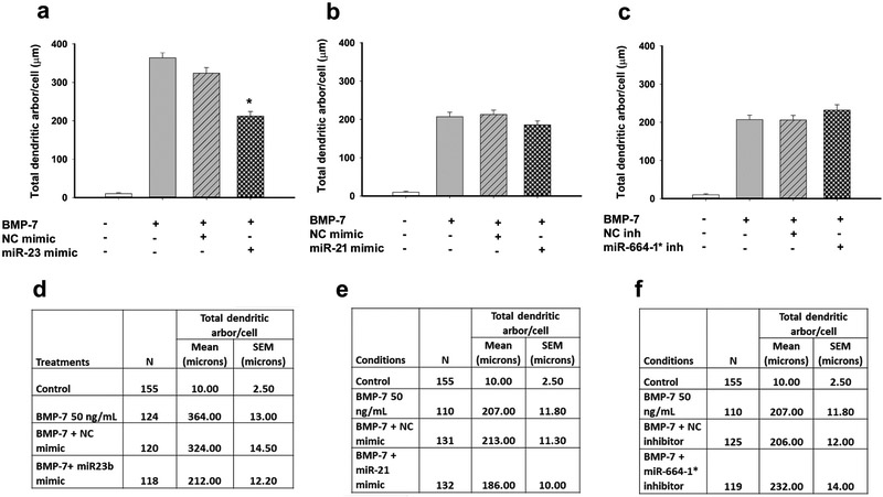
Fig. 7. Effect of miRNA modulation on total dendritic arbor of BMP-7 treated neurons.
Sympathetic neurons were transfected with mimics for miR-21 or miR-23b or inhibitors for miR-664-1* using Lipofectamine RNAimax. Controls were untransfected or transfected with negative controls (NC) for the miRNA mimics and inhibitors. Following transfection, cultures were treated with control medium or BMP-7 (50 ng/mL) for 5 d. The total length of the dendritic arbor per neuron was quantified in cultures immunostained for MAP-2 to identify dendritic processes. Data are expressed as the mean ± SEM. Panels A-C show the graphical representation of the data and D-F show the numerical values including N for each condition, mean and SEM. Statistical significance was assessed using one-way ANOVA, followed by Tukey’s post hoc test. *Significantly different from cultures transfected with respective negative controls in the presence of BMP-7 at p ≤ 0.05.

