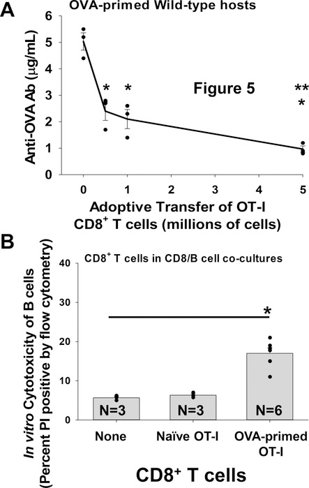
Figure 5. OVA-primed OT-I CD8+ T cells suppress anti-OVA antibody production in a dose-dependent manner and mediate in vitro cytotoxicity of OVA-primed B cells.
A) WT mice were primed with mOVA lysate (i.p. injection) on day 0. Cohorts of WT mice received adoptive transfer (AT) on day 0 of increasing quantities of OVA-primed OT-I TCR transgenic CD8+ T cells (0, 0.5, 1, 5×10⁶ cells i.v.). Mouse serum was assayed for anti-OVA antibodies (Ab) by ELISA on day 14 following antigen stimulation. WT mice primed with mOVA lysate exhibit maximal levels of serum anti-OVA antibodies on day 14 following antigen stimulation (5.0±0.3 μg/mL; n=3). AT of 0.5, 1, or 5×10⁶ OVA-primed OT-I CD8+ T cells on day 0 inhibited anti-OVA antibody production in these mice (2.4±0.4, 2.1±0.4, and 1.0±0.1 μg/mL respectively; n=3 and p<0.0004 for all groups signified by “*”). The AT of 5×10⁶ OVA-primed OT-I CD8+ T cells inhibited anti-OVA antibody production significantly more than 0.5 or 1×10⁶ OVA-primed OT-I CD8+ T cells (p<0.04 for both signified by “**”). B) OVA-primed OT-I CD8+ T cells or naïve OT-I CD8+ T cells were co-cultured with OVA-primed IgG1+ B cell targets. CD8+ T cells and B cells were co-cultured at a 10:1 ratio for 4 hours and analyzed for cytotoxicity. Significant cytotoxicity of OVA-primed IgG1+ B cells was observed in co-cultures with OVA-primed OT-I CD8+ T cells (17±1.4%; n=6, p<0.001 signified by “*”) compared to negative control cultures with no CD8+ T cells (5.7±0.3%) or with naïve OT-I CD8+ T cells (6.3±0.3%). Error bars indicate standard error.

