4.
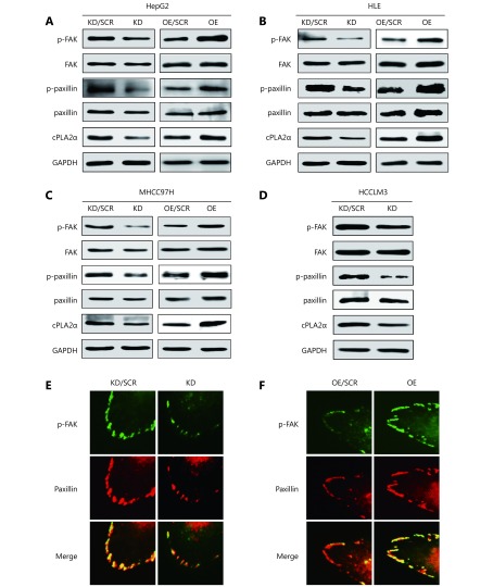
cPLA2α regulates the phosphorylation and colocalization of FAK and paxillin. (A) Western blot analysis of cPLA2α, p-FAK (Tyr-397), FAK, p-paxillin (Tyr-118) and paxillin in cPLA2α KD/SCR, KD, OE/SCR and OE HepG2 cells. (B) Western blot analysis of cPLA2α, p-FAK (Tyr-397), FAK, p-paxillin (Tyr-118) and paxillin in cPLA2α KD/SCR, KD, OE/SCR and OE HLE cells. (C) Western blot analysis of cPLA2α, p-FAK (Tyr-397), FAK, p-paxillin (Tyr-118) and paxillin in cPLA2α KD/SCR, KD, OE/SCR and OE MHCC97H cells. (D) Western blot analysis of cPLA2α, p-FAK (Tyr-397), FAK, p-paxillin (Tyr-118) and paxillin in cPLA2α KD/SCR and KD HCCLM3 cells. (E) Immunofluorescence staining of p-FAK (Tyr-397) and paxillin as well as their colocalization in cPLA2α KD/SCR and KD HepG2 cells. (F) Immunofluorescence staining of p-FAK (Tyr-397) and paxillin as well as their colocalization in cPLA2α OE/SCR and OE HepG2 cells. Original magnifications, 400 ×.

