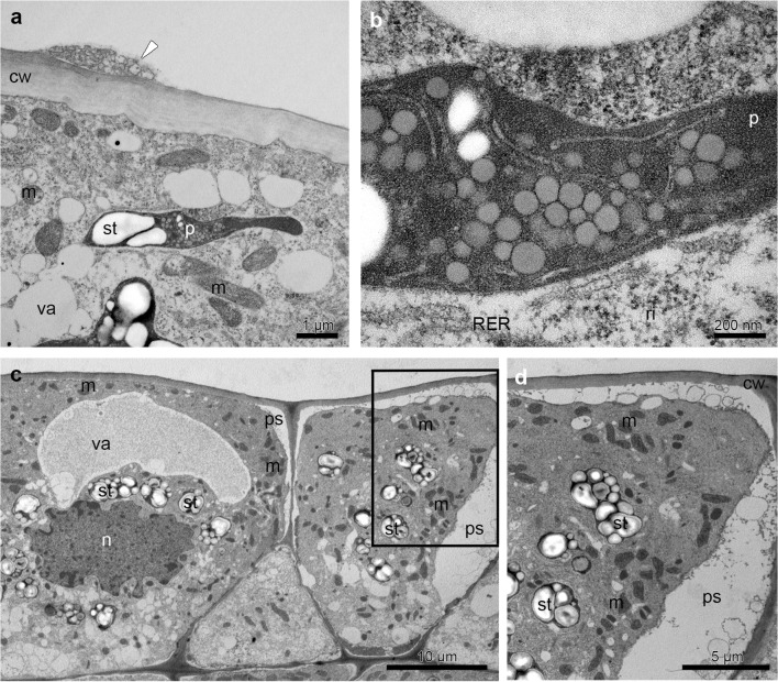
Fig. 8.
Ultrastructural analysis (TEM) of the prickles on the hypochile (a, b) and basal part of the epichile (c, d) revealed a residues of secretory material on the cuticle surface (white arrowhead), plastids with plastoglobuli, tubules and starch grains (a–d) and profiles of SER and RER (b), numerous mitochondria (a–d). c Epidermal cells containing a prominent periplasmic space with flocculent material (c, d, d detail of c), dense cytoplasm with enlarged nuclei (c). cw cell wall, d dictyosome, m mitochondrion, n nuclei, p plastid, ps periplasmic space, ri ribosomes, st starch grains, va vacuole

