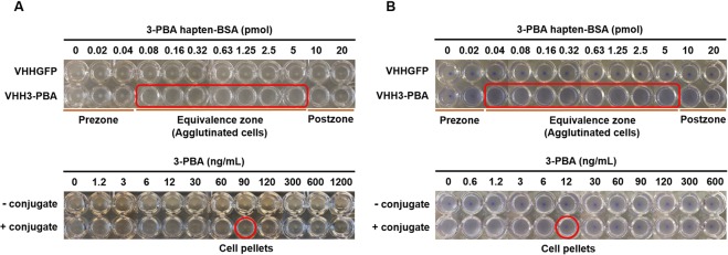
Figure 2.
Agglutination assay for 3-PBA detection in the absence (A) and presence (B) of amilCP expression. Determination of 3-PBA hapten-BSA concentration necessary to induce agglutination of E. coli DH10B cells displaying anti-3-PBA VHH (VHH3-PBA, Top) and detection of 3-PBA in response to cell pellet formation at constant concentration of the conjugate (0.08 pmol with no amilCP co-expression and 0.04 pmol with amilCP coexpression) (Bottom). Red rectangles and circles represent regions in which cell agglutination occurred and the limit of detection, respectively. Pictures are a composite of photos that were taken every six wells per frame using a phone camera. This experiment was conducted in three biological replicates.

