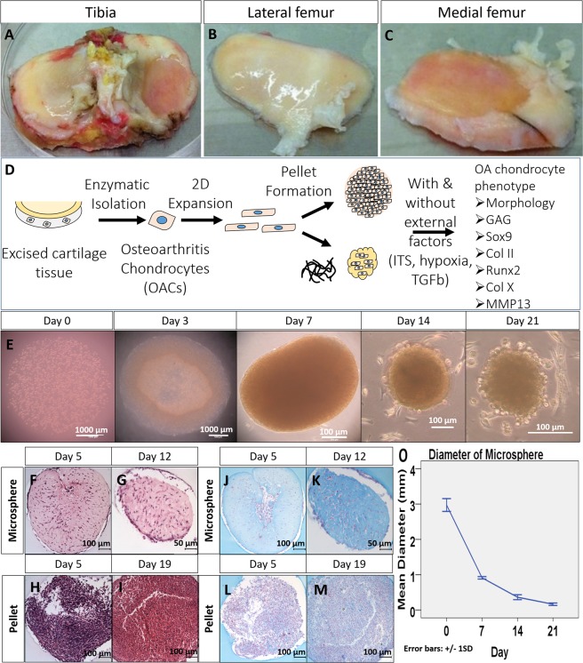
Figure 1.
General experimental design and gross appearance, H&E and Alcian blue staining of chondrocyte encapsulated collagen microspheres, compared with pellet. Excised tissues from tibia plateau (A), lateral (B) and medial femoral condyle (C) were collected from total knee replacement surgery. Schematic diagram of the overall experimental design (D). In brief, chondrocytes in the cartilage were enzymatically isolated from the cartilage specimens and expand in monolayer culture. The cells (P3) were then cultured in 3D in order to resume its in vivo characteristics. Two kinds of 3D culture method, namely pellet culture and collagen microencapsulation were used for chondrocyte 3D culture and the effect in phenotypes restoration were compared. Collagen microspheres at different time points during a 3 week culture (E). H&E staining (F–I) and Alcian blue staining (J–M) of microsphere and pellet. Diameter of microsphere during a 3 week culture (O), from 5 independent experiments, each with diameter measurement of 30 microspheres at each time points.

