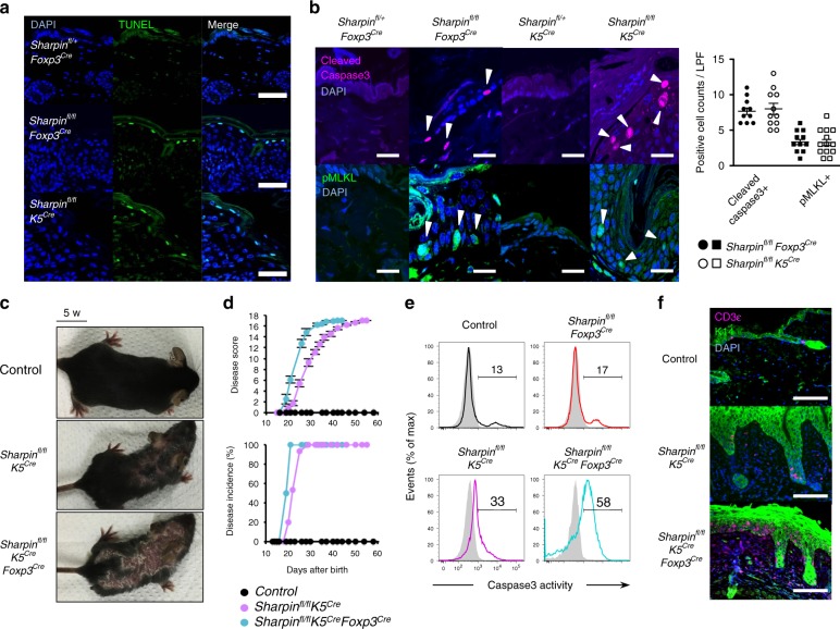
Fig. 5.
Skin-oriented T cells induce apoptotic and necroptotic keratinocyte death in an inflammatory setting. a TUNEL staining of skin sections from 5-week-old Sharpinfl/flK5Cre, 10-week-old Sharpinfl/flFoxp3Cre, and control mice. Scale bars: 50 μm. b Immunofluorescence staining of skin sections. Cleaved caspase-3 and phosphorylated MLKL were used as markers of apoptosis and necroptosis, respectively. Scale bars: 20 μm. Cell death in the epidermis was quantified by counting the total number of bright cells per low-power field (LPF). c Appearance of skin dermatitis. d Progression (upper) and incidence (lower) of skin disease among the indicated strains (numbers of mice: control, n = 12; Sharpinfl/flK5Cre, n = 15; Sharpinfl/flK5CreFoxp3Cre, n = 8). Disease scores were recorded up to 60 days after birth. No gender-based differences in the incidence of skin disease were observed. e Representative histogram showing death-prone epidermal cells from the indicated strains. Isolated epidermal cells were incubated with PhiPhiLux-G1D2, a fluorescent substrate of active caspase-3. f Immunofluorescence staining of skin sections to detect skin-infiltrating T cells. Scale bars: 100 μm. The small horizontal lines in b, d indicate the mean (±s.e.m.). Data are representative of at least three independent experiments. Source data are provided in a Source Data file

