Figure 2.
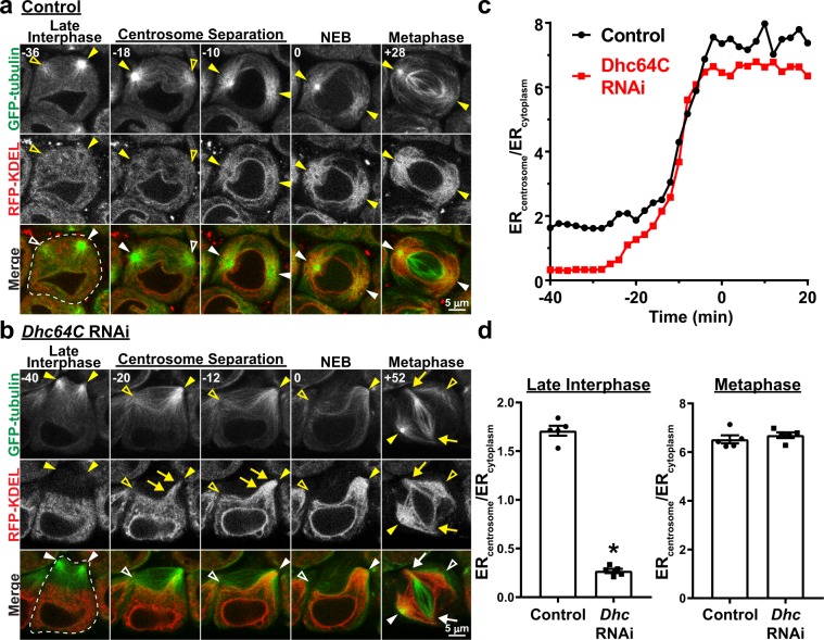
ER association with astral MTs during M-phase is independent of dynein activity. (a) Representative images of a control spermatocyte co-expressing GFP-tubulin (green) and RFP-KDEL (red) from late interphase through metaphase of the first meiotic division. Times in minutes in the upper left of each image are reported relative to NEB, which was assigned as the first timepoint at which there was a notable increase in GFP-tubulin fluorescence in the nucleus. Filled arrowheads indicate visible centrosomes, and open arrowheads indicate the approximate location of centrosomes that are out of the focal plane. (b) Representative images of a Dhc64C RNAi spermatocyte co-expressing GFP-tubulin (green) and RFP-KDEL (red) from late interphase through metaphase of the first meiotic division. Times in minutes in the upper left of each image are reported relative to NEB. Filled arrowheads indicate visible centrosomes, and open arrowheads indicate the approximate location of centrosomes that are out of the focal plane. Arrows in the “Centrosome Separation” images (−20 and −12 min) point to RFP-KDEL – labeled ER that is moving toward the centrosome along astral MTs. Arrows in the “Metaphase” image (+52 min) point to the spindle poles where the kinetochore MTs are focused; note that the centrosomes with astral MTs and associated ER are completely dissociated from the spindle poles. (c) The ratio of RFP-KDEL fluorescence intensity immediately adjacent to a single centrosome versus that in the cytoplasm near the nucleus (ERcentrosome/ERcytoplasm) was calculated every 2 minutes from late interphase (−40 min) through metaphase (20 min) for the control and Dhc64C RNAi cells shown in (a,b), respectively. Time is relative to NEB. (d) Mean ERcentrosome/ERcytoplasm ± SEM for five control and Dhc64C RNAi cells during late interphase (left panel; measured at 40 min prior to NEB) and metaphase (right panel; measured at 20 min post NEB). ‘*’, p < 0.0001, t-test.

