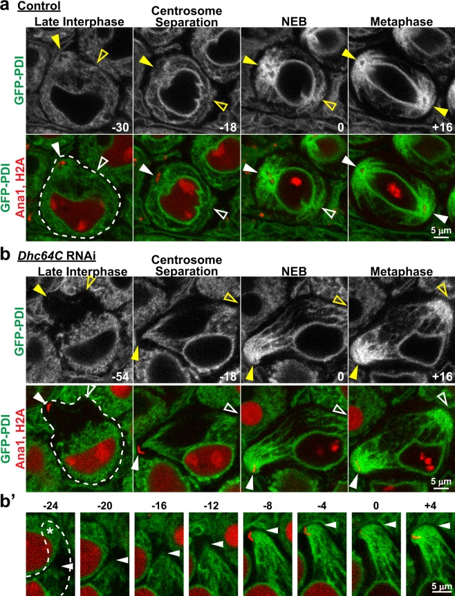
Figure 3.
Spindle pole focusing of the ER during M-phase is independent of dynein activity. (a) Representative images of a control spermatocyte co-expressing Ana1-tdTomato, H2A-RFP (red) and GFP-PDI (green) from late interphase through metaphase of the first meiotic division. Times in minutes are reported relative to NEB, which was assigned as the first timepoint at which the diffuse H2A-RFP fluorescence in the nucleus dissipated. Filled arrowheads indicate visible centrosomes marked by Ana1-tdTomato, and open arrowheads indicate the approximate location of centrosomes that are out of the focal plane. (b) Representative images of a Dhc64C RNAi spermatocyte co-expressing Ana1-tdTomato, H2A-RFP (red) and GFP-PDI (green) from late interphase through metaphase of the first meiotic division. Times in minutes are reported relative to NEB. Filled arrowheads indicate visible centrosomes, and open arrowheads indicate the approximate location of centrosomes that are out of the focal plane. (b’) Expanded timecourse of GFP-PDI being drawn toward one of the centrosomes in the Dhc64C RNAi spermatocyte. The arrowheads indicate the leading front of GFP-PDI. In the first four frames the centrosome to which GFP-PDI is being drawn is out of the plane of focus, but its approximate location is indicated by ‘*’ in the first frame.

