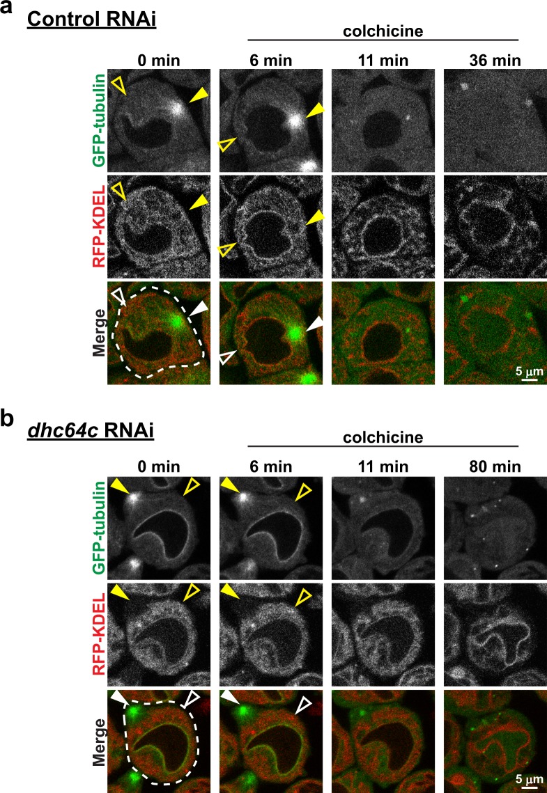
Figure 4.
Dynein independent ER redistribution to centrosomes requires MTs. Control (a) and Dhc64C (b) RNAi spermatocytes expressing GFP-tubulin (green) and RFP-KDEL (red) were imaged beginning in late interphase before the commencement of ER redistribution to the centrosomes. Colchicine (50 µM) was then applied 6 min later and the cells were imaged through NEB to encompass the time during which ER redistribution would normally occur. Note the lack of any distinct focus of the ER by the time of NEB in the control cell, and that the ER fails to be drawn to the cell cortex, where the centrosome was located prior to colchicine application, in the Dhc64C RNAi cell.

