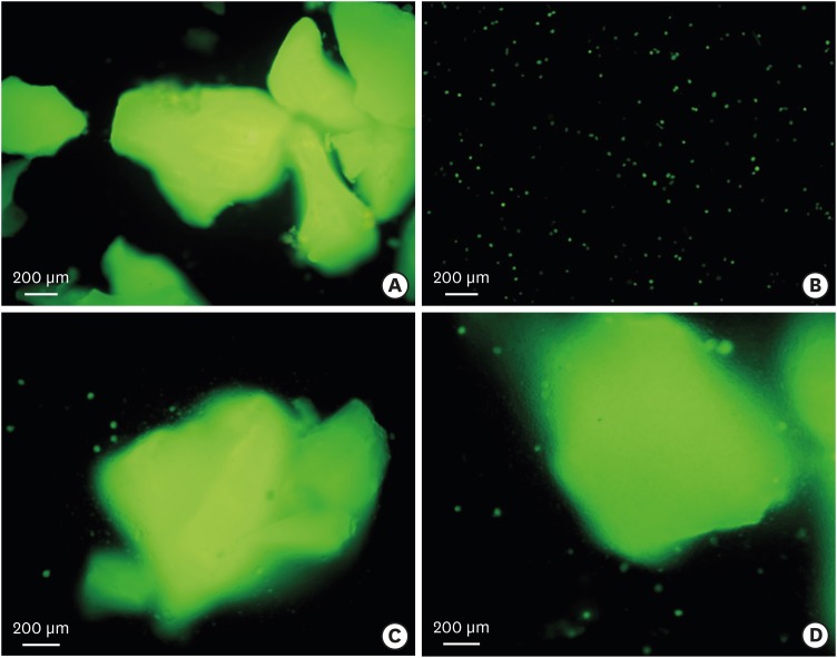
Figure 3. Fluorescent microscopic evaluation of the grafted tissue. (A) Group 1 (β-TCP/HA+MSCs): Viable MSCs were attached to β-TCP/HA. (B) Group 2 (collagen membrane+MSCs): The surviving MSCs that had been transplanted were easily separated from the collagen membrane. (C) Group 3 (β-TCP/HA+collagen membrane+MSCs). The surviving MSCs that had been transplanted were abundantly attached to the β-TCP/HA, and a small amount of MSCs were scattered around the β-TCP/HA. (D) Group 4 (β-TCP/HA+chipped collagen membrane+MSCs). The results were similar to those of group 3.
β-TCP/HA: β-tricalcium phosphate/hydroxyapatite, MSCs: mesenchymal stem cells.

