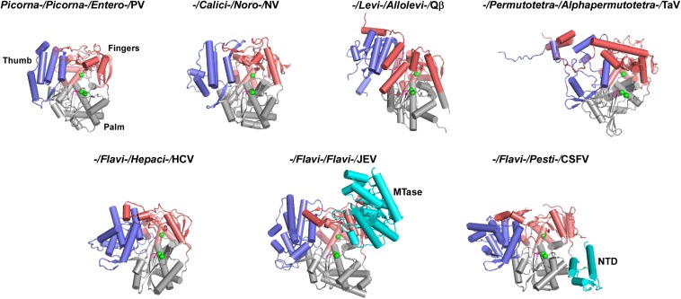
FIGURE 3.
Global views of seven representative RdRPs 3D structures. RdRP structures are shown in cartoon representations. If available, order/family/genus/species assignments are shown on top of each structure. PV, poliovirus, PDB entry 1RA6 (chain A); NV, norovirus, PDB entry 1SH0 (chain A); Qβ, bacteriophage Qβ, PDB entry 3MMP (chain G); TaV, Thosea asigna virus, PDB entry 4XHI (chain A); HCV, hepatitis C virus, PDB entry 1C2P (chain A); JEV, Japanese encephalitis virus, PDB entry 4K6M (chain A); CSFV, classical swine fever virus, PDB entry 5YF5 (chain A). Coloring scheme: RdRP palm in gray, thumb in blue, fingers in pink, and signature sequence XGDD in magenta. The α-carbon atom of the three absolutely conserved amino acid residues (labeled by asterisks in Figure 2) are shown as green spheres. The N-terminal additional regions, if present, are shown in cyan.

