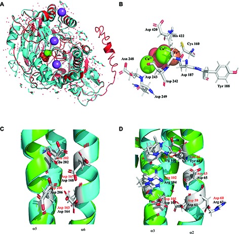
Figure 2.

Structural analysis of PhoD and PhoU of Halothece sp. PCC 7418. (A) Predicted structure of PhoD of Halothece sp. PCC 7418 is represented in red and aligned with the crystal structure 2YEQ that is displayed in blue cyan. (B) Active center of PhoD of the test bacterium showing all the aminoacids involved in coordination with two Ca2+ and Fe3+ ions. (C) Cluster 1 of PhoU, trinuclear metal site with Fe, between α5 and α6. (D) Cluster 2 of PhoU, tetranuclear metal site with three Fe and one Ni, between α2 and α3. Black amino acids are from predicted PhoU and red amino acids are from 4Q25. All the structures were represented with Pymol.
