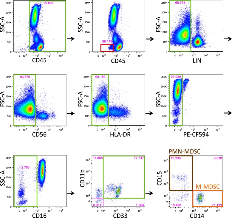
Fig. 1.
Gating strategy for identification of MDSC. Fresh whole blood (WB) samples (100 μL) served as the substrate for the WB flow cytometric assay. Green boxes indicate the cell populations that were selected for continued analysis. Red boxes indicate cell populations that were excluded. Initial exclusion of cellular doublets and debris, by gating on the singlets, is not shown. CD45+ cells were selected, followed by basophil exclusion, both using plots of CD45 vs SSC-A. Subsequently, T and B cells were excluded by gating on cells negative for pooled anti-CD3, anti-CD19 and anti-CD20 antibodies (lineage negative, LIN−) cells. NK cells were excluded by gating on CD56− cells, and HLA-DR− cells were selected. Eosinophils were excluded by gating on the PE-CF594− cell population. Neutrophils were excluded by gating on CD16− cells. Total myeloid derived suppressor cells (MDSC) were defined as CD33 + CD11b + cells. Polymorphonuclear-MDSCs (PMN-MDSC, a subset of total MDSCs, brown box) were identified by CD15+ expression, while monocytic-MDSCs (M-MDSC, a subset of total MDSCs, orange box) were identified by CD14+ expression. Early stage MDSC (e-MDSC), or counterpart MDSC in healthy individuals [7] are shown in the final plot, bottom left quadrant, as CD3/19/20/56−HLA-DR−CD16−CD33 + CD11b + CD14−CD15− cells

