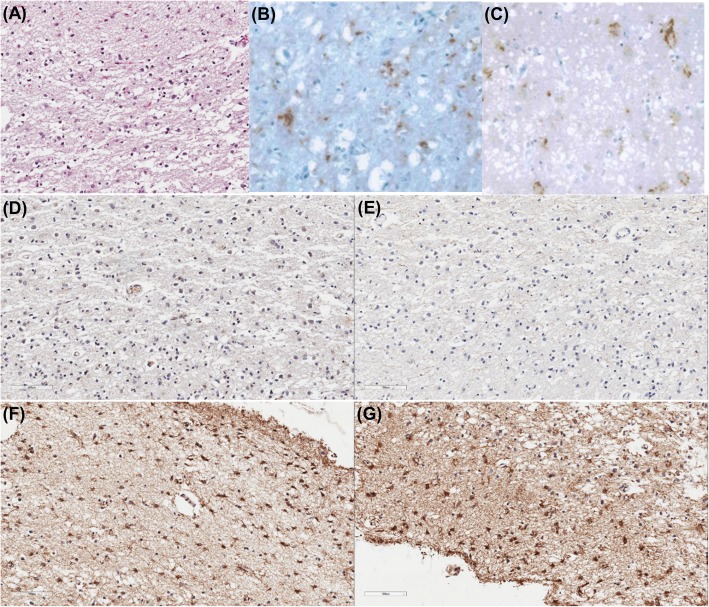
Fig. 2.
Pathological findings in the left occipital cortex of a patient with sporadic Creutzfeldt-Jakob disease. H&E staining (a) showed neuronal loss and vacuolation. Immunohistochemistry showed reactivity for PrPsc, b 3F4 antibody and c 1C5 antibody. Immunohistochemistry against amyloid-ß (d) and phosphorylated tau (e) showed no amyloid plaques and no neurofibrillary tangles, respectively. GFAP staining (f) showed active astrocytosis and MAO-B staining (g) showed increased MAO-B activity

