Fig. 1.
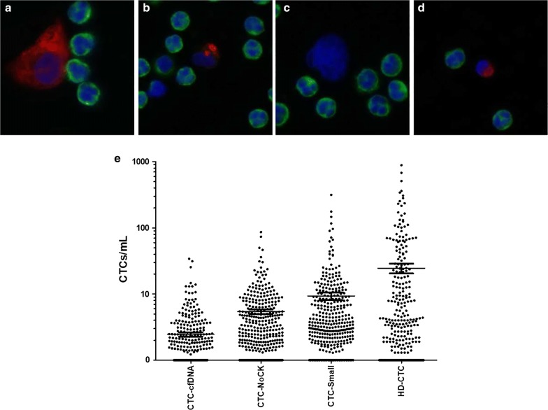
HD-CTC and candidate CTC cell data for stage IV NSCLC. a Representative image of HD-CTC. HD-CTCs are cytokeratin positive (red), CD45 negative (green), contains a DAPI nucleus (blue) and is morphologically distinct from surrounding white blood cells. b–d Representative images of types of suspected candidate CTCs found in a single NSCLC patient. b Nucleus too small and cytoplasm insufficiently circumferential; cell appears to be in late apoptosis and defined as CTC-cfDNA producing. c Suspected CTC that is negative for cytokeratin and CD45, but has a nucleus that is morphologically similar to CTCs, defined as CTC-NoCytokeratin. d Nucleus is small (same size as surrounding WBC nuclei) and cytokeratin present, defined as CTC-Small. e Distribution of CTCs and candidate cells in NSCLC patients

