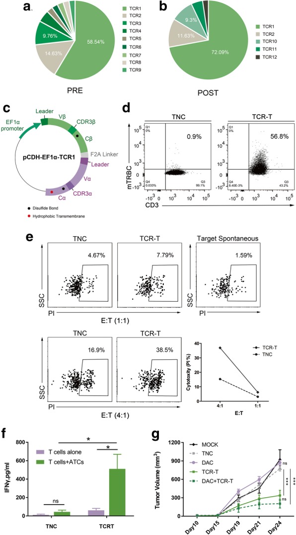
Fig. 4.

Identification of tumor-specific TCRs and functional verification of corresponding TCR-Ts. TCRs distribution of CD8+ CD137+ T cells in pre- (a) and post-stimulated (b) TIL-F1 by single-cell RT-PCR analysis. TCRs sequences are listed with different colors in order from most to least frequent, named TCR1 to TCR12, respectively. c Sketch map of pCDH-EF1α-TCR1 lentiviral vector. The construct employed the β-α chain order, added murine constant region, disulfide bond (presented as black dots), and α chain hydrophobic-substitutions (presented as red dot). Leader, leader sequences of TCRα and TCRβ chains, respectively; EF1α promoter, elongation factor 1 alpha promoter; F2A linker, Furin-P2A linker. d Transduction efficiencies were measured by staining cells with an anti-murine TCR-β chain constant region antibody. The results were representative of independent experiments done with more than three different donors. e Cytotoxicity capacity of TCR-Ts against ATCs. Line chart summarized the cytotoxicity by subtracting the ATCs spontaneous death at different E: T ratios. Data were representative of at least three independent experiments with more than three different donors. f IFN-γ ELISA measurement of TCR-Ts and TNC targeting ATCs. The results are representative of more than three independent experiments in more than three different donors (*p < 0.05, Student paried t test). g Antitumor activity of TCR-Ts against patient derived xenograft models. The tumor volume is plotted on the y axis. Time after tumor cell injection is plotted on the x axis. The mean values from each group are plotted. Error bars represent the SEM (n = 5 mice per group, ***p < 0.001, analyzed by two-way repeated measures ANOVA). The results are representative of 2 independent experiments. MOCK, none; TNC, two intravenous injections of untransduced T cells; DAC, a single intraperitoneal injection of DAC; TCR-T, two intravenous injections of TCR-Ts; DAC + TCR-T, two intravenous injections of TCR-Ts and a single intraperitoneal injection of DAC
