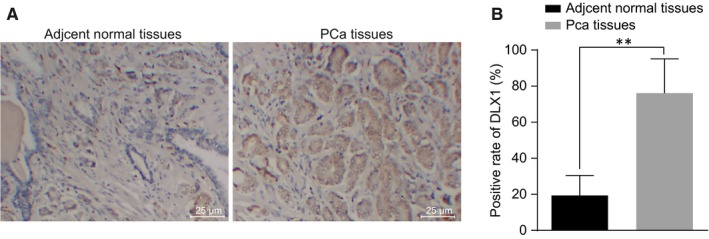
Figure 2.

The positive expression of DLX1 protein is increased in PCa tissues. A, immunohistochemical staining analysis of DLX1 protein in the PCa tissues and adjacent normal tissues; B, quantitative analysis for the positive expression rate of DLX1 protein in the PCa tissues and adjacent normal tissues; **P < 0.01, compared with PCa tissues or adjacent normal tissues; PCa, prostate cancer; DLX1, distal‐less homeobox 1
