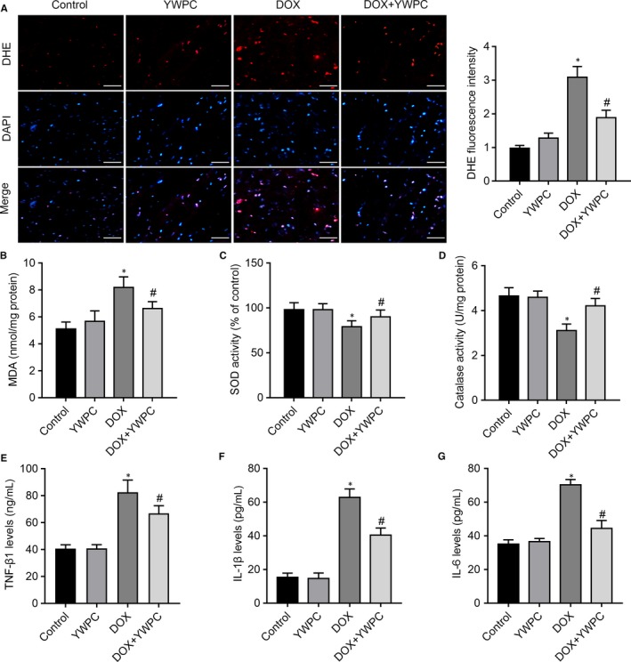
Figure 3.

YWPC treatment ameliorates DOX‐induced oxidative stress, in vivo. (A) Staining cells with red indicated DHE‐positive cells; DAPI staining (blue) indicated nucleus. Bar = 50 μm. Percentages of DHE‐positive cells of total cells were shown. Cardiac antioxidant enzyme activities of (B) MDA, (C) SOD and (D) catalase were evaluated using in the heart homogenates from indicated groups. Enzyme‐linked immunosorbent assay (ELISA) was used on serum from rats to determine the expression of transforming growth factor‐β1 (TGF‐β1) (E), interleukin‐1β (IL‐1β) (F), and interleukin‐6 (IL‐6) (G). Data are shown as the mean ± standard deviation of 10 rats. *P < 0.05, vs. the control; # P < 0.05 vs. the DOX group. DOX, doxorubicin; MDA, malondialdehyde; SOD, Superoxide Dismutase; YWPC, Yellow Wine Polyphenolic Compounds
