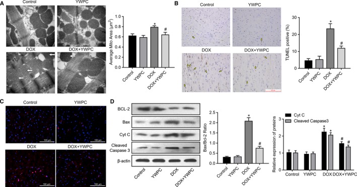
Figure 4.

YWPC prevent DOX‐induced cardiac myocyte apoptosis, in vivo. (A) Transmission electron microscopy showing betterment of mitochondrial ultrastructure in DOX + YWPC group compared to DOX‐treated rats. Bar = 0.2 μm, 50 000× magnification. Corresponding graphs showing mitochondrial diameter and average mitochondrial area in vivo from different treatment groups (n = 10). (B) Representative images of TUNEL staining of the heart tissues. Apoptotic cardiomyocyte nuclei appear brown‐stained and normal nuclei appear blue. (C) Caspase‐3 expression in the different treatment groups were detected via immunofluorescence analysis. (D) Protein expression of Bcl‐2, Bax, Cyt C and cleaved caspase‐3 in the different treatment groups. *P < 0.05, vs. the control; # P < 0.05 vs. the DOX. DOX, doxorubicin; YWPC, Yellow Wine Polyphenolic Compounds
