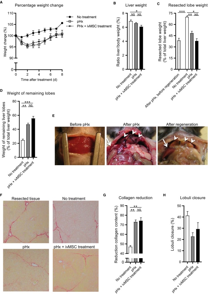
Figure 2.

Systemically administrated MSCs did not further improve the pHx initiated reversal of fibrosis. Mice with liver fibrosis received no treatment, pHx or pHx + ivMSC (N = 6/9 per group). A, Normalized body weight during regeneration. Relative weights of (B) total liver, (C) remnant parts of the resected lobes and (D) remaining lobes after regeneration. E, Pictures of the liver before pHx, after pHx and after regeneration. Remnant and remaining lobes are indicated with white arrow heads and white arrows respectively. F, Sirius‐red stained liver tissue (20x magnifications) (G) Reduction of Sirius‐red staining relative to resected tissue. H, Estimated lobuli closure. Data are expressed as mean ± SEM. *P ≤ 0.05, **P ≤ 0.01, ***P ≤ 0.001. MSCs, mesenchymal stromal cells; pHx, partial hepatectomy; NS, not significant
