
Figure 2.
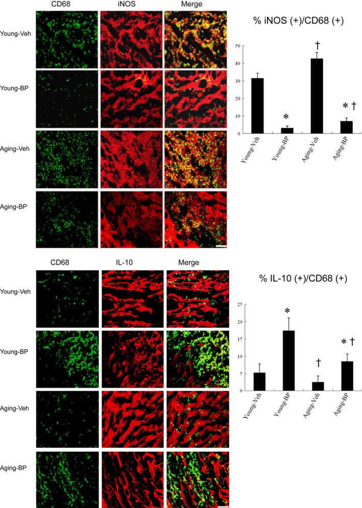
Immunohistochemical staining of M1 and M2 macrophage phenotype at the border zone at day 3 after MI. Upper panels. iNOS‐expressing CD68 (+) M1 macrophages were recruited to infarcted myocardium treated with vehicle (Veh), but were significantly reduced after n‐butylidenephthalide (BP) administration. The M1 ratio was significantly higher in ageing rats than in young rats. Lower panels. IL‐10‐expressing CD68 (+) M2 macrophages were predominant in BP‐administered infarcted myocardium. The ageing rats had a significant lower M2 ratio compared with the young rats. The iNOS‐expressing CD68 (+) or IL‐10‐expressing CD68 (+) macrophages were calculated and expressed as bar graphs. The values are mean ± SD of five animals from each group. The line length corresponds to 20 μm. *P < 0.05 vs vehicle in respective age group; † P < 0.05 vs the same treatment in the young group