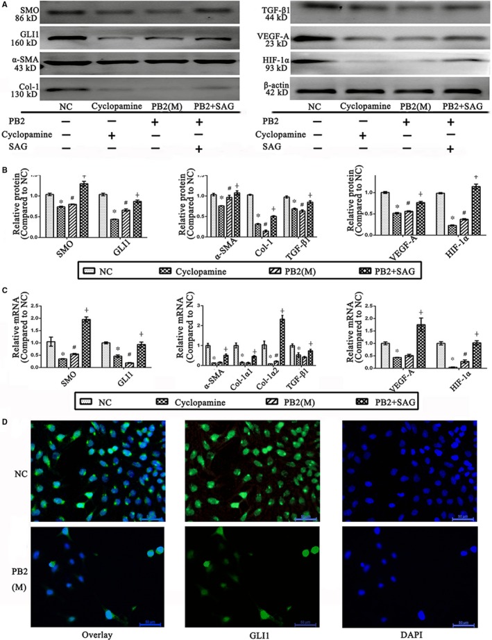
Figure 6.

Effects of PB2 on the Hh pathway in LX2 cells. A, Effects of cyclopamine, PB2, and SAG on the Hh pathway, and the activation and angiogenesis ability of LX2 cells. B, The quantitative analysis of Western blot results. C, The levels of SMO, GLI1, α‐SMA, Col‐1α1, Col‐1α2, TGF‐β1, VEGF‐A and HIF‐1α mRNA in LX2 cells. D, IF staining of GLI1 in LX2 cells (original magnification, 400×). All of the experiments were repeated in triplicate. Data are presented as the mean ± SD from three independent experiments. * Indicates P < .05 vs the NC group; # indicates P < .05 vs the L group; and ┼ indicates P < .05 vs the M group
