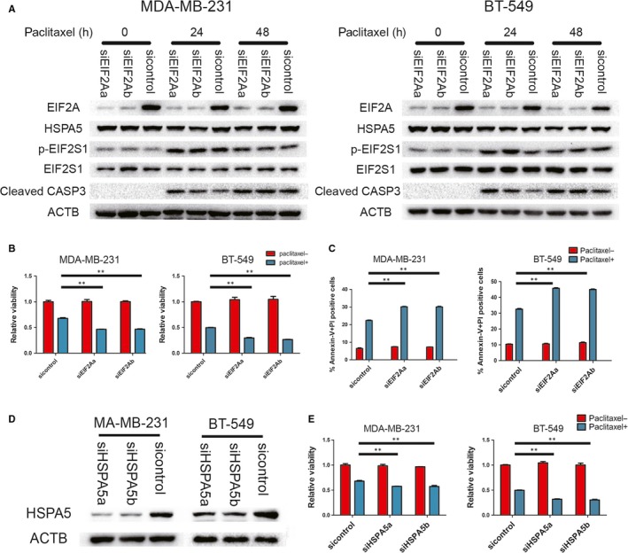
Figure 6.

EIF2A promotes cell survival during paclitaxel treatment in vitro. A, Indicated cells were transfected with EIF2A siRNA, and followed by paclitaxel treatment. Cells were lysed at indicated time points for Western blot assays of EIF2A, HSPA5, p‐EIF2S1, EIF2S1 and cleaved caspase 3. B and E, Indicated cells transfected with indicated siRNA were incubated with 100 nmol/L paclitaxel for 48 h. Cell viability was measured by CCK‐8 assay. Percentage of cell survival is represented as mean ± SD from 3 independent experiments (n = 3, mean ± SD). **P < 0.01, Student's t test. C, indicated cells stained with Annexin‐V and PI, and analysed by FACS. Bars indicate mean values ± SD of three experiments. **P < 0.01. D, MDA‐MB‐231 and BT‐549 transfected with indicated siRNA. Western blots were performed with indicated antibodies
