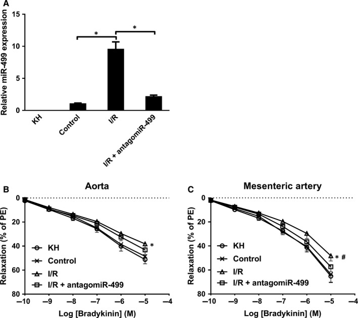
Figure 6.

Perfusate from I/R impairs endothelium‐dependent relaxation. A, The amount of miR‐499 was significantly higher in I/R compared to control perfusate, non‐detectable in KH, and significantly decreased by addition of antagomiR‐499 (n = 3 and P < 0.05 for all). B, The I/R perfusate (△) decreased ~21% and ~26% of EDR at 10−5 and 10−6 M of Bradykinin, respectively, relative to control (×) and KH buffer (○) in aorta (n = 3 and P < 0.05), and addition of antagomiR‐499 (□) tends to restore EDR (n = 3 and P > 0.05). C, The I/R (△) decreased ~25%, ~27% and ~29% of EDR at 10−5 10−6, and 10−7 M of Bradykinin, respectively, relative to control (×) and KH buffer (○) in mesenteric artery (n = 3 and P < 0.05), and antagomiR‐499 (□) abrogated the miR‐499‐mdiated decrease of EDR (#compared to I/R, n = 3 and P < 0.05). Experiments were performed three times in triplicate. Data are presented as ‘Mean ± SEM’. Dose‐response curves were analysed with 2‐way analysis of variance, with * denoting P < 0.05 and # P < 0.05.
