
Figure 3.
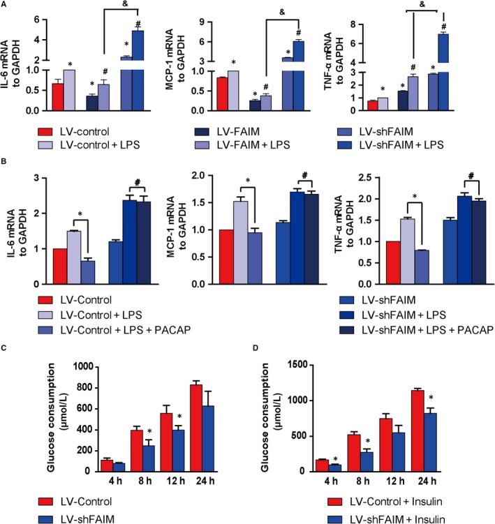
Effects of FAIM on inflammatory cytokine production and glucose metabolism. A, Cells were cultured for 24 h and then were stimulated with 0.1 g/mL LPS for 6 h. The expression levels of IL‐6, MCP‐1 and TNF‐α were analysed by real‐time PCR. *P < 0.01, groups versus LV‐Control group; #P < 0.01, groups versus LV‐Control + LPS group; &P < 0.01, LV‐shFAIM + LPS group versus LV‐FAIM + LPS group. B, PACAP was added to rescue the effects of FAIM knockdown, and then the mRNA expression levels of pro‐inflammatory factors were detected. *P < 0.01, PACAP + LPS groups versus LPS groups; #P > 0.05, PACAP + LPS groups versus LPS groups. C, Accumulated glucose consumption of AML12 cells. The AML12 cells were cultured in 6‐well plates, the glucose levels in the medium were detected at 4, 8, 12 and 24 h, and the cumulative glucose consumption was calculated. *P < 0.01, LV‐shFAIM groups versus LV‐Control groups. D, After cells were treated with insulin (100 nM), the cumulative glucose consumption was calculated at 4, 8, 12 and 24 h. *P < 0.01, LV‐shFAIM + insulin groups versus LV‐Control + insulin groups. Data are presented as mean ± SEM