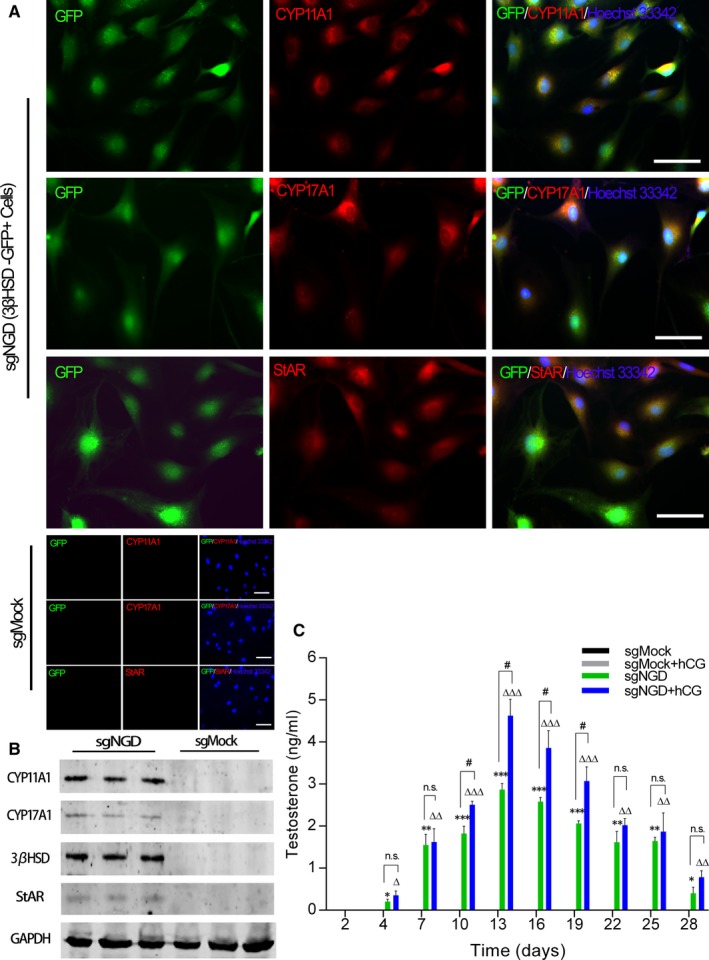
Figure 4.

Further characterization of Hsd3b‐GFP+ cells. A, Immunofluorescence staining revealed the expression of steroidogenic enzymes at day 14 postinfection of the sgNGD lentivirus. Hoechst 33342 was used to counterstain nuclei (blue). Scale bars = 50 µm. B, Representative Western blotting results for the protein expression of Leydig steroidogenic markers in induced Leydig‐like cells at day 14 postinfection with the sgNGD lentivirus. C, Time course of testosterone production during culture after infection with the sgRNA lentivirus. Every 3 days, the testosterone concentration in the supernatant was measured, and the supernatant was replaced by fresh medium either containing hCG (10 ng/mL) or not. *P < 0.05, **P < 0.01, ***P < 0.001, significant difference compared to sgMock; △ P < 0.05, △△ P < 0.01, △△△ P < 0.001, significant difference compared to sgMock + hCG; # P < 0.05, significant difference between the indicated groups; ns, not significant. Data are presented as the mean ± SD of 3 biological replicates
