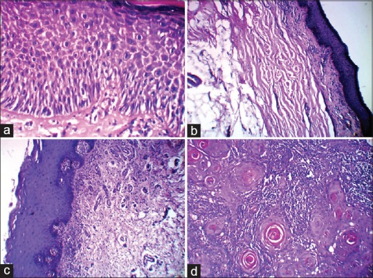
Figure 3.

The photomicrograph showing hyperkeratinised mucosa with moderate epithelial dysplasia as in leukoplakia (a). Atrophic epithelium and dense collagen fiber bundles in oral submucous fibrosis (b). Basal cell degeneration and juxta-epithelial inflammatory infiltrate as in lichen planus (c). And epithelial cells in the form of islands, sheets, cords and strands with numerous keratin pearl formation as in well differentiated squamous cell carcinoma (d). (H&E, ×40)
