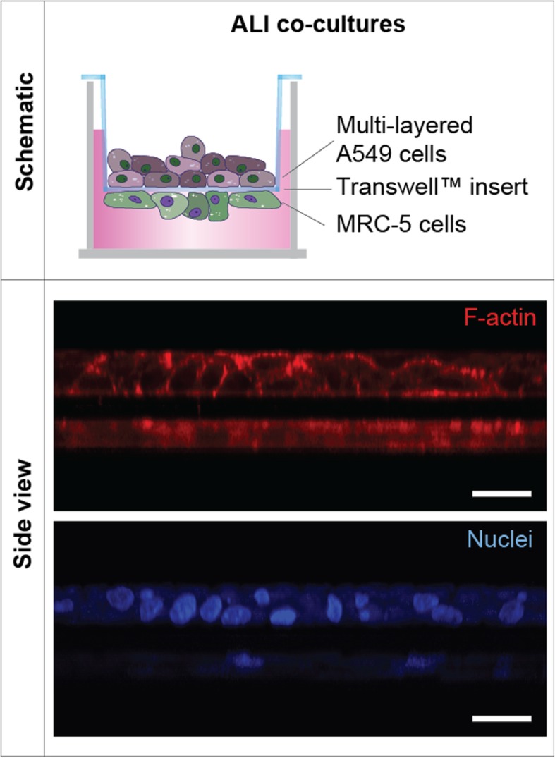
Fig. 1.

Composition and structure of ALI multilayered co-cultures. Schematics of the in vitro model developed and representative LSCM images of the culture showing the F-actin (in red) and cell nuclei (in blue) organization in these cultures. The Z-stack LSCM images, clearly demonstrating the 3D architecture of the models developed, were reconstructed with ImageJ software to obtain the side view shown. Scale bars: 20 μm (objective lens, 63×)
