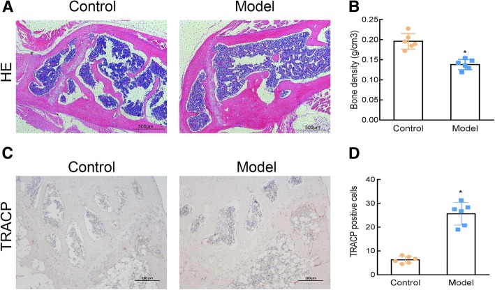
Fig. 2.
Osteoporosis (OP) was induced in mice with retinoic acid (RA). HE staining (40X) (a), femur bone mineral density determination (b), and TRACP staining (100X) (c) were performed to investigate the changes in femoral trabecular structure, bone density, and number of osteoclasts (d) in the OP model. Control: normal mice, Model: RA-induced OP model mice. *, P < 0.05 vs control; n = 6 per group

