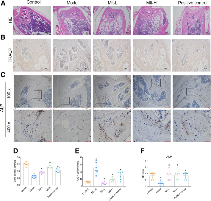
Fig. 6.
Melatonin improves the femur trabecular bone and reduces the number of osteoclasts of osteoporosis (OP) model mice. After the administration of melatonin or alendronate to trabecular bone for 2 months, distal femur was detected by HE staining (40X) (a), TRACP staining (100X) (b), and ALP IHC staining (100X and 400X) (c). Bone density was assessed (d), and the number of TRACP-stained osteoclasts was quantified (e). The IHC staining intensity was scored (f). Control: normal mice, Model: retinoic acid (RA)-induced OP model mice, Mlt-L: low-dose melatonin-treated OP model mice, Mlt-H: high-dose melatonin-treated OP model mice. Positive control: alendronate-treated OP mice. *, P < 0.05 vs control. #, P < 0.05 vs model; n = 6 per group

