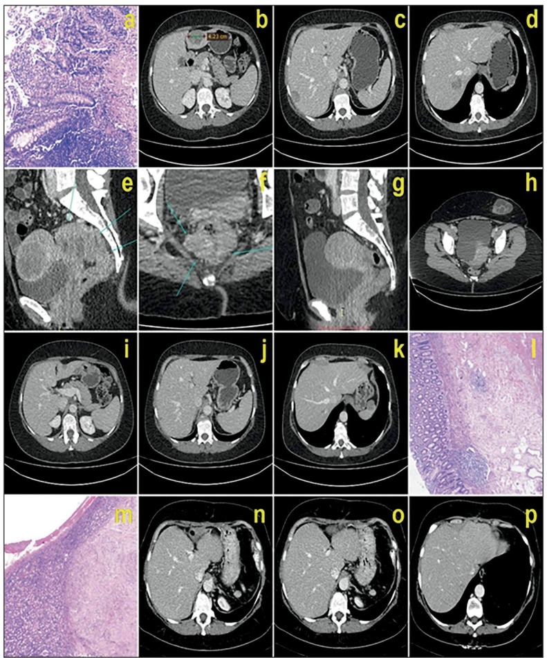
Figure 1.

(a): Preoperative histopathology-adenocarcinoma; (b), (c), (d), (e), (f): preoperative CT of primary and secondary tumors; (g), (h), (i), (j), (k): CT after neoadjuvant therapy; (1), (m): complete pathological response of primary rectal tumor and secondary liver metastases in S3, respectively; (n), (o), (p): CT shows vanishing liver metastases 4 months after the operation.
