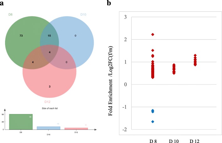
Fig. 8.
Venn diagram for X-linked differentially expressed genes (a) and expression profile of X-linked differentially expressed genes (b). Green, blue, and pink filed in Venn graph indicate X-linked differentially expressed genes on Day 8, 10, and 12 of the embryos, respectively. The red and blue plots in the scatter mean the up and down-regulated genes on the X chromosome in female embryos, respectively

