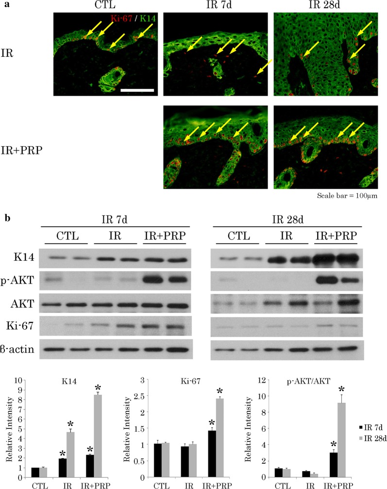
Fig. 5.
Proliferation of K14-positive cells in radiation skin-injury mouse model. a Ki-67 positivity (arrows) on immunostain; and b Western blot analysis of p-AKT, AKT, and Ki-67 expression levels at wound sites. Results of one experiment performed in triplicate and expressed as mean ± SEM *p < 0.05 vs. IR group. IR irradiation, CTL control, PRP platelet-rich plasma

