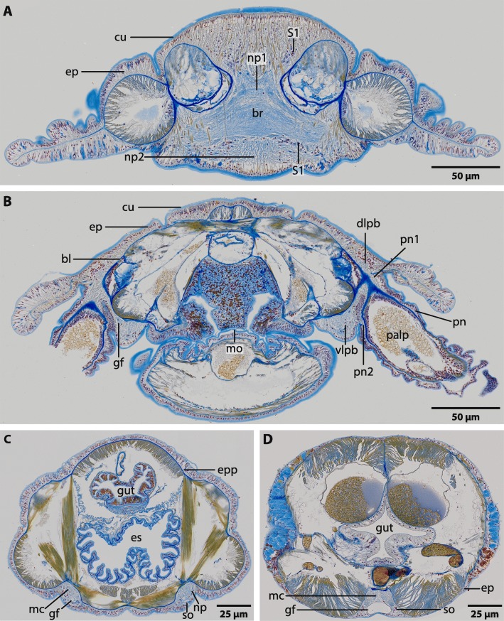
Fig. 2.
Magelona mirabilis, histological cross sections (5 μm), Azan staining, frontal (a) to caudal (d). a: The brain (br) of M. mirabilis is located inside the epidermis (ep). Frontally it is composed of two different types of neuropil (np1, np2). Neurites in np1 are arranged in parallel to the body axis, while neurites in np2 are arranged longitudinal to the body axis. Somata of the first type (S1) are located ventro- and dorso- laterally of the neuropil. cu: cuticle. b: Posteriorly the brain is composed of a dorsal and a ventral lobe. Both, the dorso- lateral (dlpb) and the ventro- lateral parts of the brain (vlpb) give rise to two nerves (pn1, pn2) which connect the palp nerve (pn) to the brain. bl: basal lamina; gf: giant fibre; mo: mouth opening. c: The brain gives rise to two laterally located medullary cords (mc). Somata (so) are located ventrally to the neuropil (np). An epidermal plexus (epp) is present and surrounds the whole animal. Both giant fibres (gf) can be traced to the ventro-lateral part of the brain. es: esophagus. d: Both medullary cords fuse ventrally to a single cord (mc) which is located in an epidermal invagination. Both giant fibres also fuse to a prominent single fibre (gf). Somata (so) are located ventro- laterally to the neuropil

